

Treatment depends on severity. Mild cases may need only monitoring, while children whose lids block the pupil often need correction to protect normal visual development.
At Kopelman Aesthetic Surgery, Dr. Joel E. Kopelman evaluates congenital ptosis with a focus on both function and natural-looking eyelid balance.
Key Takeaways
- Congenital ptosis is a drooping upper eyelid present at birth, seen in about 1 in 842 live births. Most cases (76%) are simple congenital ptosis, caused by an underdeveloped eyelid-lifting muscle.
- Doctors grade severity by how far the lid sits over the pupil (MRD1, normally 4 to 4.5 mm). A drop of 2 mm or less is mild, about 3 mm is moderate, and 4 mm or more is severe.
- The biggest risk is to your child’s vision, not appearance. About 1 in 7 affected children develop amblyopia (lazy eye), and about 1 in 5 develop strabismus; amblyopia may not be reversible after ages 7 to 10.
- Levator muscle function decides the surgical approach. If the muscle is 5 mm or more, surgeons can shorten it (resection); if it is 4 mm or less, they use a sling. Most repairs happen between ages 3 and 5, sooner if the lid blocks vision.
- Surgery does not always last. Reported revision rates range from about 20% in one review to 50% or more over 8 to 10 years in another, so plan on long-term follow-up with a specialist.
Understanding Congenital Ptosis Meaning
What Is Ptosis (Congenital Lid or Palpebral Ptosis)?
Ptosis is the medical term for a drooping eyelid. This drooping eyelid medical term comes from the Greek for “falling,” and doctors also use the ptosis medical term blepharoptosis [1][3].
Congenital lid ptosis, also known as congenital blepharoptosis, occurs when a child is born with a low upper lid or develops one during the first year. It can affect one or both eyes and varies in severity.
Simple congenital ptosis occurs in about 1 in 842 live births, according to a 40-year Minnesota population study [4].
The condition is usually painless but can interfere with vision and development. Early diagnosis and proper management help prevent long-term problems such as lazy eye (amblyopia).
Types and Severity of Eyelid Droop
Congenital ptosis is graded by how far the eyelid blocks the visual field. Doctors measure the distance from the lid margin to the pupil’s light reflex, called MRD1. A normal reading is about 4-4.5 mm [1].
Grades reflect the MRD1 difference between the two eyes, or the difference from normal when both sides are affected [1][3].
| Grade | MRD1 difference (vs. other eye or normal) | What you may notice | Usual next step |
|---|---|---|---|
| Mild ptosis | 2 mm or less | Slight droop; the pupil stays clear and vision is not impaired | Monitoring; optional correction before school age |
| Moderate ptosis | About 3 mm | Lid edge partially covers the pupil; brow raising or head tilt | Close vision checks; surgery if vision, astigmatism or posture is affected |
| Severe ptosis | 4 mm or more | Lid edge reaches the pupil center or lower; chin-up posture is common | Surgical correction, often earlier to protect vision |
Mild ptosis rarely affects sight. It may still be worth treating before school age if the eyes’ difference is easy to see.
Knowing the grade helps guide the right ptosis treatment in NYC, especially in young patients.
Is Congenital Ptosis a Disability?
Congenital ptosis is not classified as a disability in most cases, but severe forms can affect vision and learning. When the droop limits daily function, treatment becomes medically necessary rather than cosmetic.
Children may need school accommodations if vision is compromised.
Congenital vs. Acquired Eyelid Ptosis
Congenital ptosis is present at birth or within the first year. Acquired ptosis develops later due to aging, trauma, or medical conditions; because the causes and treatment paths differ, an accurate diagnosis matters.
One clinical clue helps distinguish the two. A congenital droop often sits higher when the child looks down, because the stiff muscle can’t relax. An acquired droop from a stretched levator tendon usually stays low in downgaze [1].
Ptosis is also different from extra lid skin. The medical term for saggy eyelids caused by loose skin is dermatochalasis, which blepharoplasty treats rather than ptosis repair [1]. A careful evaluation separates these conditions.
When One Eyelid Droops: Causes of Ptosis in One Eye
When one eyelid droops from birth, simple congenital ptosis is the most likely reason. Most congenital cases affect only one side [1][3].
Other causes of ptosis in one eye in children include neurological or syndromic conditions (see the types table below) and a mass or swelling in the lid, such as a hemangioma or plexiform neurofibroma [1][2].
What causes droopy eyelids in adults? Common causes include age-related stretching of the levator tendon, long-term contact lens wear, eye surgery, injury, and nerve or muscle disorders such as myasthenia gravis [1].
Ptosis Causes and Risk Factors
Muscle, Nerve, and Genetic Droopy Eyelid Causes
Parents often call ptosis a lazy eyelid, and lazy eyelid causes fall into three groups: muscle, nerve, and genetic. The most common is poor development of the levator muscle, the main muscle that lifts the lid. This is different from lazy eye (amblyopia).
In the affected muscle, fat and fibrous tissue replace normal fibers, so it can’t fully contract or relax [1]. That is why the lid droops at rest and lags in downgaze.
Neurological causes of ptosis are less common. Abnormal nerve signals from the brain to the lid cause the droop in conditions such as third cranial nerve palsy, Horner syndrome, and Marcus Gunn jaw-winking syndrome [1][2].
Genetic syndromes such as BPES, linked to the FOXL2 gene, can also contribute [1]. The table under Types of Congenital Ptosis compares them.
Most cases are isolated, but family history and underlying syndromes raise the likelihood.
Risk Factors in Newborns and Infants
Congenital ptosis may appear as part of a broader syndrome or occur on its own. Risk factors include:
- A family history of ptosis or related conditions
- A genetic syndrome such as BPES
- Congenital cranial nerve palsy
- Birth trauma, including forceps-assisted delivery [1]

Types of Congenital Ptosis and How They Differ
Simple congenital ptosis is the most common type, making up about 76% of all childhood ptosis cases in one population study [4]. The other types come with eye, nerve, or muscle findings that change the evaluation and the surgical plan.
| Type | What you may notice | Usual approach |
|---|---|---|
| Simple congenital ptosis | Weak or absent lid crease, lid lag in downgaze, reduced levator function; usually one side | Levator resection or frontalis sling, based on levator function |
| Marcus Gunn jaw-winking | Lid lifts when the child chews or moves the jaw; often noticed during feeding | Mild cases may be watched; a strong wink can need the levator released and a sling placed |
| BPES (blepharophimosis syndrome) | Severe ptosis in both eyes, narrow lid openings, skin folds at the inner corners, wide-set inner corners | Ptosis is treated first with a frontalis sling to protect vision; other lid features follow later; a genetics referral may help |
| Third cranial nerve palsy | Ptosis with limited eye movement; the pupil is often enlarged | Poor levator function; a sling raises the risk of poor lid closure and double vision, so care is shared with a strabismus specialist |
| Horner syndrome | Mild droop, smaller pupil on the same side, less sweating on that side of the face, lighter iris | Doctors look for an underlying cause, sometimes with imaging; a Müller muscle repair often fits |
| Double elevator palsy (monocular elevation deficiency) | Ptosis or apparent ptosis with limited upward gaze and a chin-up posture | Managed mainly by a strabismus surgeon; ptosis repair can follow, but lid closure may be poor |
| Rare muscle conditions (CFEOM, CPEO) | Ptosis with limited eye movement in most directions; CPEO worsens over time | Team care with a strabismus specialist; a heart check when a mitochondrial condition such as Kearns-Sayre is suspected |
A child with any type other than simple congenital ptosis often needs input from more than one specialist, such as a neurologist or geneticist [1][2][3].
Ptosis Symptoms, Signs, and Diagnosis
Early Signs in Infants and Children
Parents often notice that one or both lids droop more than usual shortly after birth. Other signs include:
- Tilting the head backward or lifting the chin to see
- Raising the eyebrows to pull the lid up
- Delayed visual tracking
- Uneven appearance of the eyes
Children with ptosis may also show visual fatigue or rely on head postures to widen their field of vision. These signs may point to congenital ptosis and should prompt an exam with a pediatric ophthalmologist or oculoplastic surgeon.
What Does Ptosis Look Like in a Child?
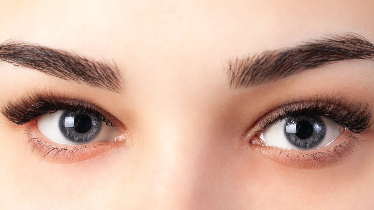
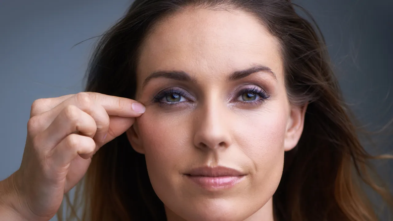
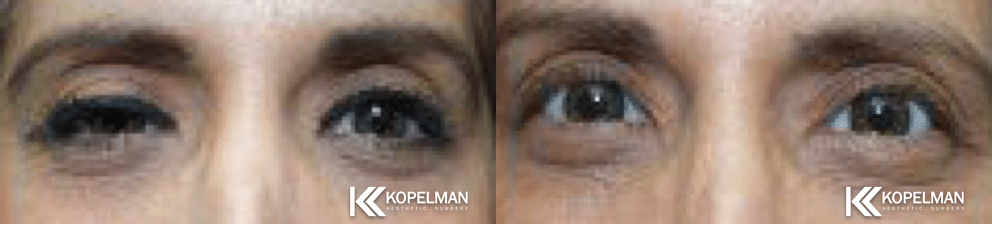
What does ptosis look like day to day? The upper lid sits lower than normal, so one eye looks smaller or sleepier. Straight-on photos make the difference easy to compare.
One sign is specific to congenital ptosis: when the child looks down, the droopy lid sits higher than the normal one. Head tilt, brow raising, and the missing crease are covered in the sections above and below.
The ptosis photos below show common presentations. The eyelid ptosis before-and-after gallery shows how lid position changes after repair. At home, dated photos taken at regular intervals help the doctor track changes over time [1].
Congenital Eyelid Crease and Visual Impact
In children with congenital ptosis, the eyelid crease may be poorly defined or absent. This is a key physical exam finding.
If the lid blocks part of the pupil, it can interfere with visual development and lead to amblyopia. A heavy lid can also press on the cornea, causing astigmatism.
In a population-based study of childhood ptosis, amblyopia affected about 1 in 7 children [5] and strabismus (eye misalignment) about 1 in 5 [6].
Some children also have refractive errors or unequal focusing between the eyes, which increases the risk of amblyopia [3]. Timing matters, since amblyopia may not be reversible after age 7 to 10 [2].
Exams, Imaging, and Functional Tests
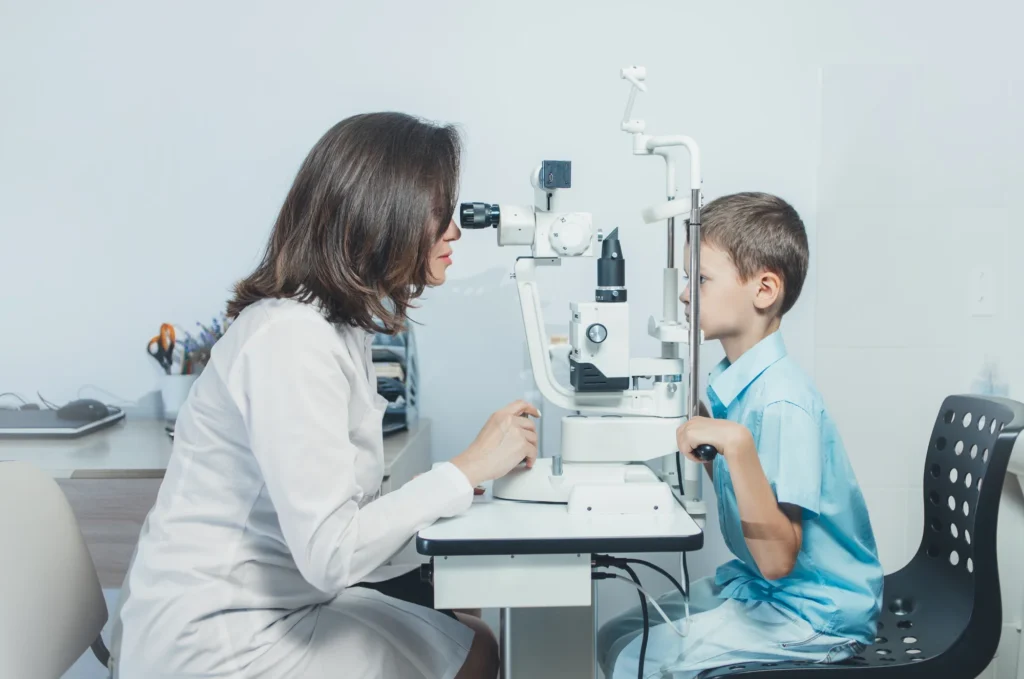
Diagnosis involves a detailed eye exam, lid-function testing, and lid-height measurements. The key number is levator function: how far the lid travels from full downgaze to full upgaze while the brow is held still. On one commonly used scale, 14 mm or more is normal, and 4 mm or less is poor [3].
The doctor also checks vision, refraction, eye alignment, and pupils. They test Bell’s phenomenon, the reflex that rolls the eye upward during lid closure and protects the cornea after surgery [1].
Doctors also rule out pseudoptosis, where a lid only looks low because the eye sits deeper, the other eye bulges, or the opposite lid is pulled up by eyelid retraction, not because the lid muscle is weak [1][3].
In some cases, imaging such as MRI is needed to assess the levator muscle or detect neurological causes. Pupil size and iris color can point to Horner syndrome [1].
Exam results determine which procedure fits. Different ptosis surgery techniques suit different levels of levator function.
Treatment Options for Congenital Ptosis
Non-Surgical Treatment Options (Including Eye Drops)
In mild cases, treatment may not be necessary, especially if vision is not affected. Some children benefit from:
- Vision monitoring every 3 to 12 months, depending on severity [2]
- Patching the unaffected eye to strengthen vision
- Corrective lenses for astigmatism or other refractive issues
- A ptosis crutch, a small bar attached to glasses that props the lid up; it is rarely used in children today and serves only as a short-term bridge [1]
Eye drops may help certain eye conditions, but they do not improve congenital ptosis. Non-surgical approaches manage symptoms rather than correct the underlying issue.
When Is Surgery Recommended?
Surgery is considered when the lid blocks the visual axis, causes amblyopia or astigmatism, or leads to a fixed chin-up head posture. In moderate to severe cases, delaying treatment may result in long-term visual impairment. Dr. Kopelman carefully evaluates each patient’s condition and growth before recommending surgery.
Correction is often necessary not only for appearance but also to prevent amblyopia and ocular torticollis, a fixed head tilt that can delay mobility in infants [2].
Even when vision is safe, some families choose repair before school age, when classmates start noticing the asymmetry [3].
Ideal Age and Surgical Techniques for Ptosis of Eyelid
Most surgical correction happens between ages 3 and 5 [1][2][3]. By then, the lid and facial structures have developed enough for accurate measurements. Severe droop in newborns, or severe droop in both eyes, may need earlier repair to protect vision [1].
The surgeon chooses a technique based on levator function [3]:
- Levator resection: Shortens the weak muscle to raise the lid; used when levator function is 5 mm or more.
- Frontalis sling: Connecting the lid to the forehead muscle so it lifts with brow movement, used when levator function is 4 mm or less.
- Müller muscle-conjunctival resection: An inner-lid approach for mild ptosis with near-normal function, used less often in children.
| Levator function | Grade | Typical procedure |
|---|---|---|
| 8 mm or more | Good to normal | Levator resection or advancement; Müller muscle-conjunctival resection for mild ptosis |
| 5–7 mm | Fair | Levator resection through the lid crease |
| 4 mm or less | Poor | Frontalis sling using fascia lata, silicone or synthetic material |
Sling materials include the child’s own fascia lata, a strip of connective tissue from the thigh that becomes usable around age 3 to 4. Surgeons also use silicone rods and synthetic sutures. Silicone is now popular because it is easy to adjust or remove [2][3].
One Lid Affected: Single Sling or Both Sides?
Both approaches work, and the choice trades symmetry against operating on a healthy lid [1][3].
A sling on the affected side alone gives acceptable symmetry when looking straight ahead. The difference shows more in downgaze and tends to shrink as the child grows [3].
Some surgeons place slings on both sides, with or without releasing the healthy lid’s muscle. Heights match more closely, but the normal lid has to be operated on, and many parents resist that [1][3].
Many surgeons choose a single sling because it delivers satisfactory results while leaving the healthy lid untouched. Bilateral surgery remains an option later, once the child can participate in the decision [1][3].
What Are the Risks of Congenital Ptosis Surgery?
The most common risks are undercorrection, where the lid stays lower than planned, and recurrence, where it drops again as the child grows. Overcorrection is rare in children [3].
| Risk | What it means | How it is handled |
|---|---|---|
| Undercorrection or recurrence | The lid stays lower than planned or drops again with growth | Repeat surgery, sometimes years later |
| Swelling, bruising and early asymmetry | Expected after surgery; early swelling can make the two sides look uneven for the first days | Settles with healing; the surgeon checks lid height at early follow-up visits |
| Incomplete lid closure and a dry cornea | The eye cannot fully close, especially during sleep, which can irritate the cornea | Lubricating ointment and close follow-up; surgeons screen for dry eye beforehand |
| Sling problems | A small lump (granuloma), infection, or material that slips, breaks, or works loose | Granulomas are usually treated conservatively and tend to resolve; a failed sling may need repeat surgery |
| Infection | Infection of the incision, or spread to the tissue around the eye | Antibiotic ointment for about a week, plus oral antibiotics after sling surgery |
| The other lid drops | After one-sided surgery, the healthy lid can droop once the operated lid lifts | Surgeons test for it before surgery by lifting the droopy lid by hand and watching the other side |
To lower these risks, surgeons check Bell’s phenomenon, tear production, and corneal sensation before surgery. They take extra care with children who have dry eyes or facial nerve weakness [1][2].
Recovery, Results, and Costs
Recovery time varies by procedure but usually involves mild swelling and bruising for 1 to 2 weeks. Most children return to school within 3 to 4 days, and mild discomfort typically responds to acetaminophen [1].
Typical care follows one published protocol, and your surgeon’s instructions come first [1][2]:
- Antibiotic ointment on the incision for about a week
- Oral antibiotics after sling surgery, if prescribed
- Lubricating ointment at naps and sleep for the first few weeks
- Patching, if the eye doctor prescribes it
- Early check visits, often every 2 to 4 weeks at first
After surgery, many children sleep with their eyes slightly open. This is expected, and the gap usually narrows over time [1].
Incisions sit in the natural lid crease, or are very small for slings. Pink scars usually fade to barely visible within a few months [1].
Patients typically see improved lid position and a more symmetrical appearance. Children sometimes need a second procedure as they grow, or when a temporary sling is replaced [1].
Reported revision rates run from about 20% in one review [3] to 50% or more over 8 to 10 years in another [2].
The cost of congenital ptosis surgery depends on the technique used, case complexity, and insurance coverage. When the droop blocks vision or risks amblyopia, an insurer may treat the repair as medically necessary. Coverage depends on the plan and on documented exam findings.
The ptosis operation cost guide explains what drives pricing for adults. A child’s plan can differ, so ask for a written estimate at the consultation.
The guide on getting insurance to pay for eyelid surgery explains the paperwork.
Living with Congenital Eye Ptosis
Emotional, Social, and Developmental Effects
Beyond vision challenges, congenital ptosis can affect a child’s confidence and social interactions. A visible droop may lead to self-consciousness or teasing, especially at school. Timely treatment can improve both appearance and self-esteem.

Long-Term Outlook and Follow-Up Care
With proper management, most children with congenital ptosis lead normal lives with minimal visual limitations. Checkups every 6 to 9 months after early visits confirm that vision continues to develop and that new issues are caught early [1].
When to Consult a Specialist
Parents should consult an oculoplastic surgeon or pediatric ophthalmologist if they notice persistent lid drooping, abnormal head posture, or visual delays.
A droop that appears suddenly or changes during the day needs prompt attention, since it can signal a neurological or muscle condition [2].
A swollen, red, or crusted lid is a different problem, such as an infection or allergy, and needs its own evaluation. The guide to common eyelid problems covers these [3].
Bring dated photos and any family history to the first visit [1]. Schedule a personalized consultation now.
Frequently Asked Questions
Is congenital ptosis genetic?
Sometimes. In a population study, about 12% of the patients asked reported a family history of ptosis [4]. Syndromes such as BPES are inherited in an autosomal dominant pattern, so one affected parent can pass the condition on [1].
Will congenital ptosis get worse over time?
Congenital ptosis can change as a child grows, so doctors recheck lid height and head posture at follow-up visits [2]. A chin-up posture that develops over time may warrant surgery [2].
References
- Shukla UV, Patel BC. Congenital Ptosis. StatPearls, NCBI Bookshelf.
- Congenital Blepharoptosis (Ptosis). EyeWiki, American Academy of Ophthalmology.
- Kratky V. Treatment of congenital ptosis. Annals of Eye Science, 2020.
- Griepentrog GJ, et al. Incidence and Demographics of Childhood Ptosis. Ophthalmology, 2011.
- Griepentrog GJ, et al. Amblyopia in Childhood Eyelid Ptosis. Am J Ophthalmol, 2013.
- Griepentrog GJ, Mohney BG. Strabismus in Childhood Eyelid Ptosis.