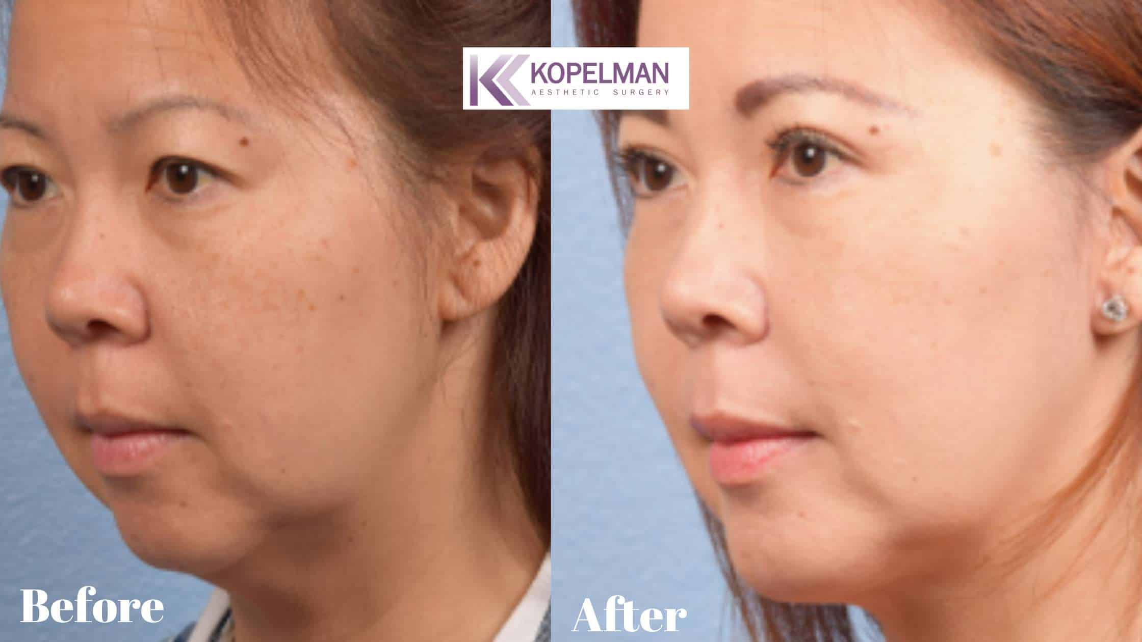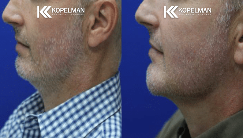
A detailed eyelid diagram is more than a picture. It helps explain how the eyelids work, why they are important, and how doctors treat them. At Kopelman Aesthetic Surgery, Dr. Joel E. Kopelman uses this knowledge to offer expert care and natural-looking results.
Understanding Eyelid Anatomy
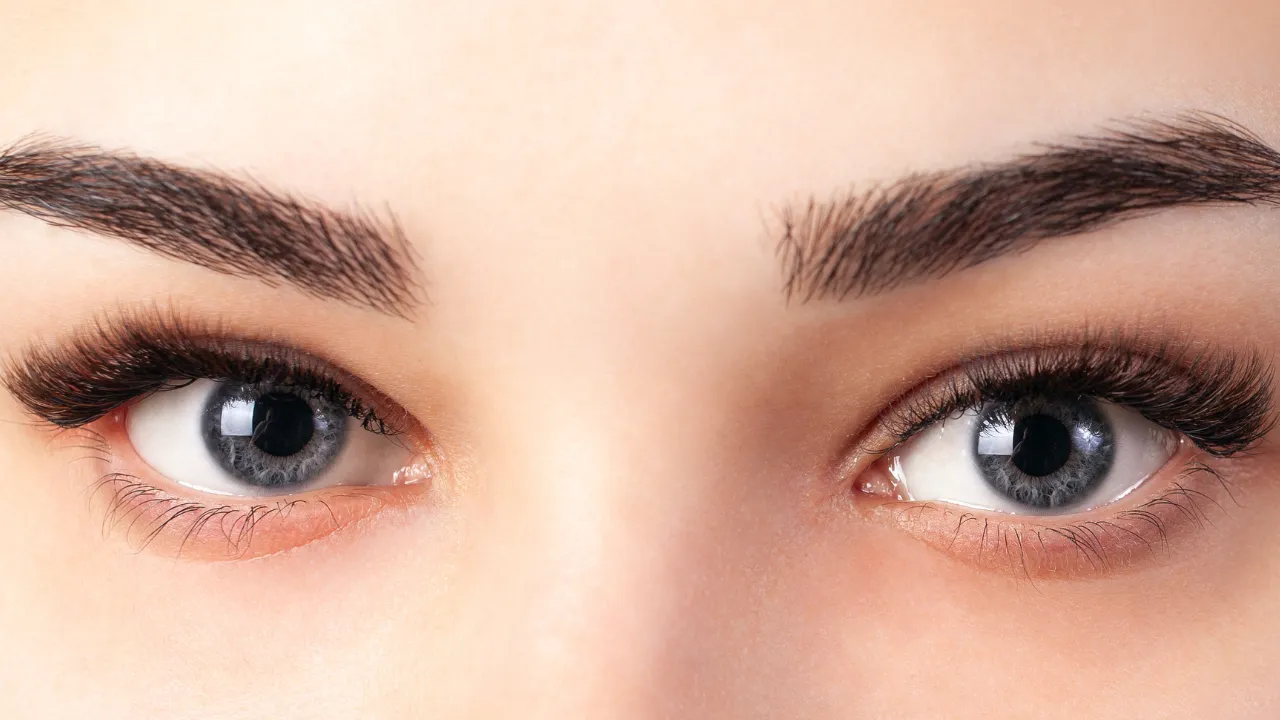
Parts of the Eyelid and Their Roles
The eyelid is made of several parts that protect the eye and help it stay healthy. These include the upper and lower lids, eyelashes, eyelid margins, and corners of the eye. Each part has a job to keep the eye safe and working well. Eyelashes prevent dust and particles from entering the eye. The eyelid margin has tiny openings that release oil to keep the eyes moist. These openings secrete an oily substance that prevents tears from drying too quickly. All these parts work together to support eye health.Eyelid Layers and Tarsal Structure
The eyelid has different layers. Knowing these helps us understand how it works. The main layers are:- Skin and subcutaneous tissue
- Muscle layer
- Firm tissue (called the eyelid tarsus)
- Conjunctiva (the inner lining)
Lower Eyelid Anatomy Overview
The lower eyelid looks like the upper lid but has some differences. It has a thinner tarsal plate and helps drain tears. Muscles in the lower lid pull it down during blinking and smiling. Supporting structures like the palpebral ligaments and the medial palpebral ligament help anchor the eyelid to the bones around the eye, including the orbital margin. One important bony landmark near the inside of the eye socket is the anterior lacrimal crest, which helps protect the tear drainage system. Behind the orbital septum are eyelid fat pads, which cushion the eye and support eyelid shape. Understanding this part is very important in surgery. Small changes here can affect how the eyelid looks and works.Eyelid Structures in Detail
Bulbar and Palpebral Conjunctiva
The conjunctiva is a thin, clear layer that covers the inside of the eyelid and the white part of the eye. The part inside the lid is the palpebral conjunctiva. The part covering the eye is the bulbar conjunctiva. It helps with tear flow and protects the eye from germs. Problems in this area, like scarring or irritation, can cause discomfort and affect eye movement.Eyelid Glands: Meibomian & Zeis
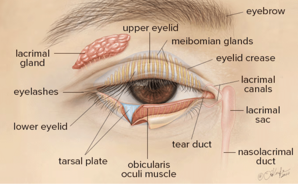
Glands in the eyelid make oils that stop tears from drying too fast. The Meibomian glands are inside the tarsal plate. They release oil to keep the tear film stable. The Zeis glands are near the eyelashes and also help with moisture.Key Eyelid Muscles and Movements
The eyelid moves because of a few key muscles. The orbicularis oculi closes the eye, and the levator muscle lifts the upper eyelid. This muscle is also known as the levator palpebra superioris (LPS). Müller’s muscle helps raise the upper lid a little more. These muscles help us blink, protect the eye, and show emotion. If they don’t work properly, they can cause droopy lids or twitching.Histology of the Eyelid Layers
Looking at the eyelid under a microscope reveals its detailed parts. The skin on the outside is thin, and the inner layer, the conjunctiva, is smooth and moist. The blood supply to the eyelid comes from branches of the internal and external carotid arteries. One key vessel is the superficial temporal artery, which supplies blood to the upper face and scalp. The nerves supply comes from sensory and motor nerves that help control movement and feeling. This level of detail is essential for surgeries like blepharoplasty. It helps the surgeon avoid damage and give better results.Common Misinterpretations in Eyelid Anatomy
Some parts of eyelid anatomy are easy to mix up. For example, people sometimes think the conjunctiva is the white part of the eye. Others think the eyelid tarsus is a muscle, but it’s actually firm tissue. To avoid mistakes:- The bulbar conjunctiva covers the white part of the eye.
- The tarsal plate is not a muscle.
- Some glands are deep inside and can’t be seen.
Labeled Diagrams and Practical Use
Labeled Eyelid Diagrams for Education & Reference
A labeled eyelid diagram shows each part of the outer eyelid clearly. It can include the eyelid layers, tarsal plate, conjunctiva, and muscles. Doctors use these diagrams to explain surgery or medical issues, and students use them to learn. They are useful tools for both.Eyelid Diagram for Makeup Placement
Knowing eyelid anatomy helps when applying makeup. A simple eyelid diagram can show where it’s safe to apply eyeshadow or eyeliner. This helps avoid skin irritation or eye problems. It’s especially helpful after surgery or for people with sensitive skin.How to Interpret an Eyelid Structure Diagram
To read an eyelid diagram:- Find the tarsal plate (firm tissue)
- Locate the muscles that move the eyelid
- Spot the conjunctiva that keeps the eye moist
- Identify where the oil glands are
- Locate the tear ducts, which help drain tears into the nose
Clinical and Surgical Relevance
Common Eyelid Conditions and Symptoms
Knowing eyelid anatomy helps doctors treat problems like:- Blepharitis: red, swollen eyelids
- Chalazion: blocked oil gland
- Entropion or Ectropion: eyelid turns in or out