If swelling or changes around the eyes are a concern, issues such as under-eye filler swelling months later should be discussed during your medical evaluation. You can ask for a short break if your eyes feel tired or dry, and you should mention symptoms such as irritation, drooping eyelids, or blurry vision before testing begins.
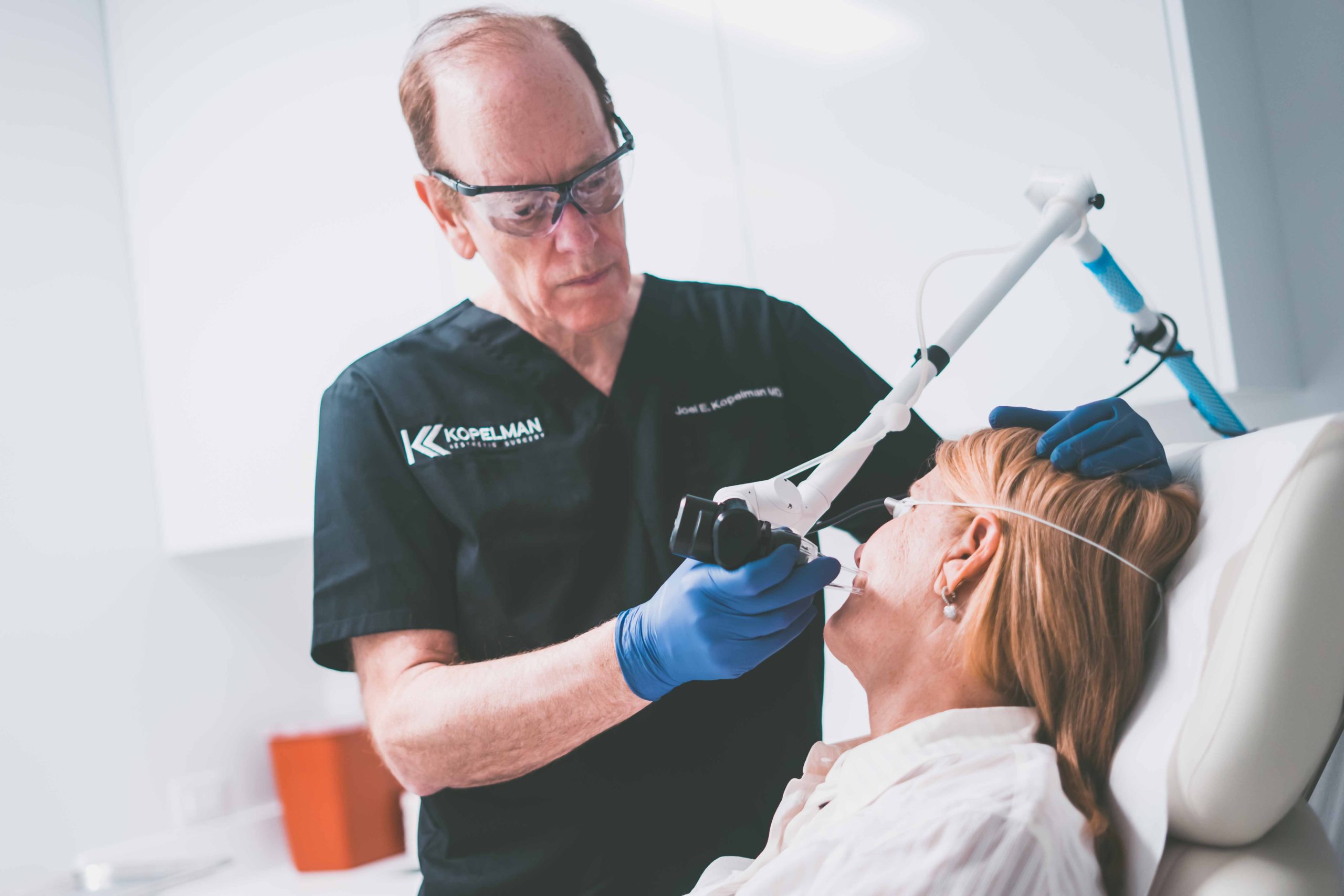
Dr. Joel Kopelman, an oculoplastic surgeon at Kopelman Aesthetic Surgery, may use visual field testing as part of a broader evaluation when assessing functional vision concerns involving the eyelids, surrounding eye structures, or symptoms such as involuntary eye closure.
Key Takeaways
- A visual field test works best when you stay focused on the center target and press the button only when you see a light.
- Missing some lights is normal because the exam uses dim targets to measure different parts of your vision.
- Good preparation includes getting enough rest, blinking normally, asking questions before the test starts, and telling the technician about eye symptoms.
- Test results may show normal vision, unreliable responses, or patterns linked to eyelid obstruction, glaucoma, optic nerve issues, or other medical concerns.
- Home checks can help you notice changes, but they cannot replace formal visual field testing in a medical setting.
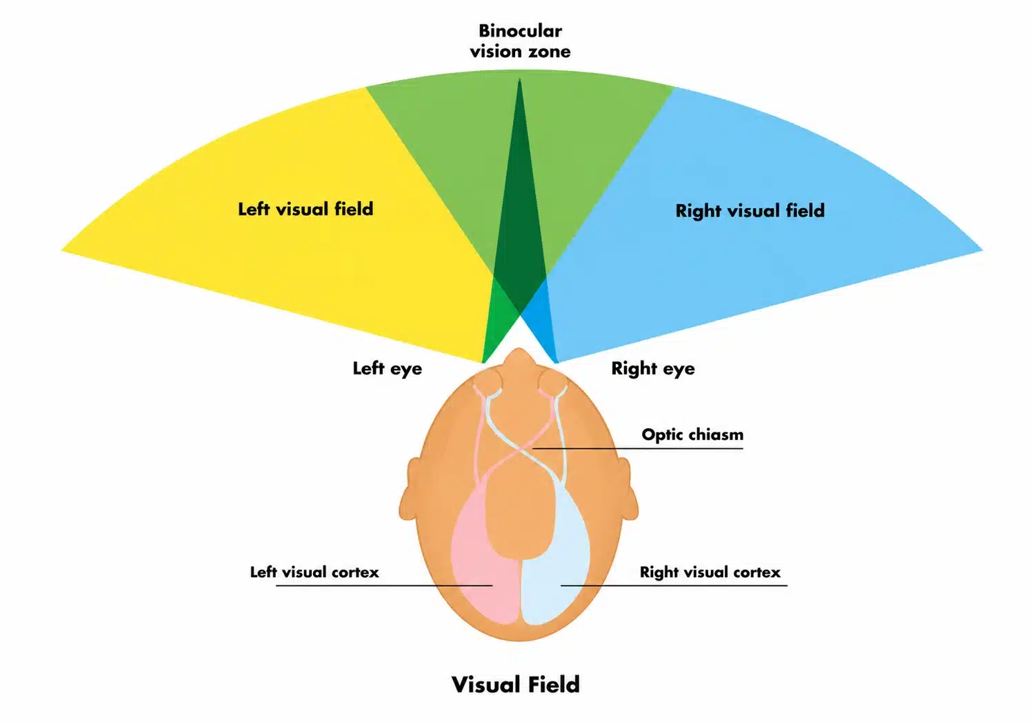
What Is a Visual Field Test?
A visual field test checks how much you can see while looking straight ahead. It measures central and peripheral vision, as well as areas where vision may be reduced or absent. Doctors use this information to detect patterns that may not appear during a standard eye chart exam.
How Do You Pass a Visual Field Test?
You do not pass this visual test by guessing, memorizing, or trying to beat the machine. The best approach is to follow instructions, stay focused, and respond only when you truly see the target. A reliable result helps your doctor understand your actual field of vision.
What the Test Measures
The test evaluates the central region, side vision, and areas near the blind spot. It can detect visual field defects caused by eye disease, eyelid obstruction, optic nerve damage, or neurological problems. It does not replace visual acuity testing, which measures how clearly you see letters or objects.
Types of Visual Field Tests
Common types of tests include:
- Automated perimetry: Measures visual sensitivity with computerized light targets.
- Humphrey visual field: A common automated perimetry test, often used for glaucoma monitoring.
- Goldmann perimetry: Uses manually controlled light targets to map the visual field.
- Tangent screen testing: Checks vision on a flat screen at a set distance.
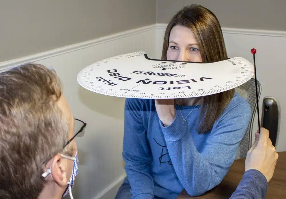
- Confrontation visual field exam: A basic screening method performed in front of the patient.
These tests help doctors measure how the eye, optic nerve, and nervous system process visual information.
Why Doctors Order Visual Field Testing
Doctors order this exam to understand the full range of a patient’s vision. The test can show whether vision loss follows a pattern linked to the eye, optic nerve, brain, or eyelids. Dr. Kopelman may consider this information when assessing functional concerns around the eyes.
Glaucoma Monitoring
Glaucoma can damage the optic nerve and reduce side vision before a patient notices symptoms. A field test can help track whether vision loss is stable or changing. Comparing past and current results can help show whether the condition appears stable or progressing.
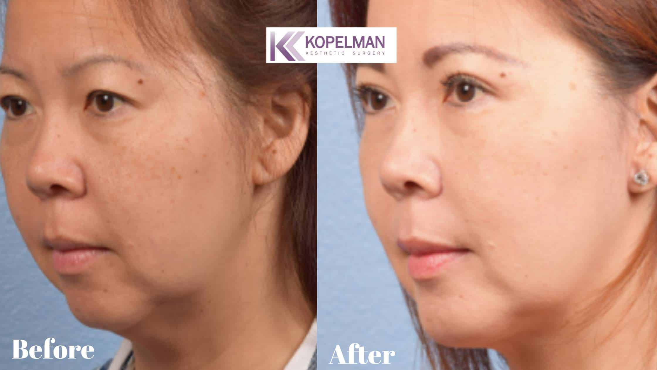
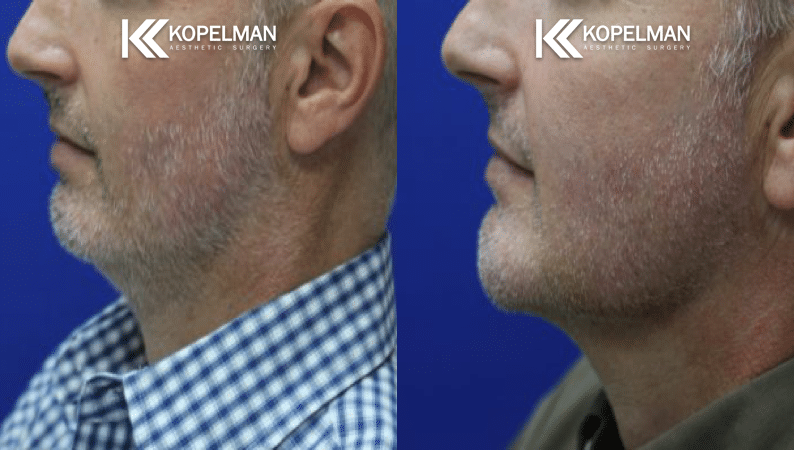
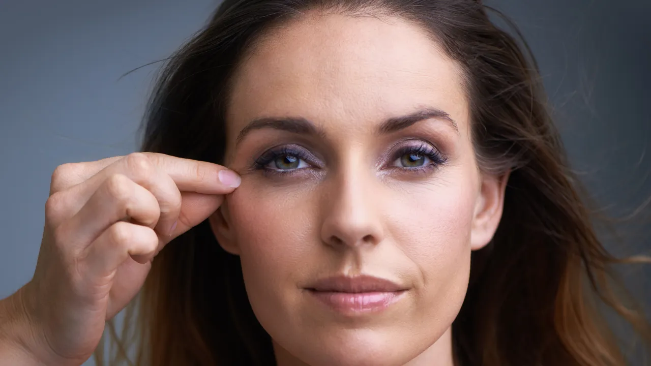
Eyelid and Vision Obstruction
Heavy upper eyelids, including cases related to aging eyelids, can sometimes block the upper part of the field of vision. In some patients, eyelid position may be affected by conditions such as congenital eyelid ptosis, which can limit the upper field of vision. This information may help distinguish cosmetic concerns from functional vision obstruction.
Visual Field Exam Tips Before Testing
Good preparation can make the exam easier and help produce more reliable results. Simple habits before the appointment may reduce eye strain, improve focus, and lower the chance of repeated testing.
- Get enough sleep the night before so you can stay focused during the exam.
- Blink normally before and during testing to help prevent dry eyes.
- Bring your glasses or contact lens information if the office requests it.
- When possible, avoid rushing into the testing room directly from bright sunlight.
- Tell the technician about blurry vision, drooping eyelids, dry eyes, headaches, or recent eye surgery.
- Ask questions before the exam starts if the instructions feel unclear.
- Take a short break during the test if your eyes feel strained or tired.
What to Tell the Technician
Tell the technician if you slept poorly, feel unwell, feel anxious, or have trouble understanding the instructions. You should also mention dry eyes, eye irritation, neck discomfort, recent eye surgery, eyelid surgery, or new vision changes. These details help the testing team adjust the setup and interpret the result with more context.
Do You Wear Glasses for the Test?
Many patients wear corrective lenses during the exam, but the testing center will guide you. The technician may place a trial lens in the machine instead of using your regular glasses. This helps test the patient under conditions that match the exam’s purpose.
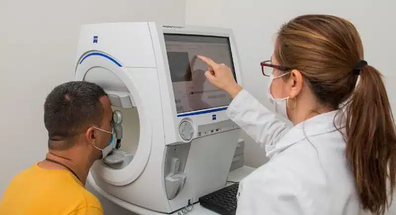
What to Do During the Test
During the exam, you sit in front of the patient testing bowl or screen and look at a fixed target. Small lights appear in different areas, and you press a button when you see one. The goal is not speed but steady, honest responses.
Keep Looking Straight Ahead
You should keep your gaze on the central target throughout the exam. Looking around the machine can make the results less reliable. If your eye moves often, the test may not reflect your true field of vision.
Press Only When You See Light
Press the button only when you actually see a light. Guessing can create false responses and make the report harder to interpret. It is normal to miss some lights because the test includes dim targets.
Ask for a Break
Ask for a break if your eyes feel strained or your attention drops. A short pause is better than forcing yourself through the test while unfocused. The technician can usually restart or continue the exam after you rest.
Why You Might Fail the Test
A poor test result does not always mean permanent vision loss, and patients who need functional eyelid evaluation may benefit from understanding how to fail an eye test for eyelid surgery in a medically appropriate context.
Some results reflect fatigue, poor fixation, dry eyes, misunderstanding, or discomfort during the exam. Other results may show a true disease that needs medical review.
Common Testing Errors
Looking around the machine, guessing too often, or trying to memorize the test can make results less reliable. You cannot memorize this exam in a useful way because targets vary by program and intensity. A field exam measures responses across space, not memorized letters on a chart.
True Vision Field Loss
Sometimes an abnormal result reflects a real loss of vision in certain areas. This can occur from glaucoma, retinal disease, optic nerve disease, stroke, or other medical causes. Your doctor reviews the pattern to decide whether more testing is needed.
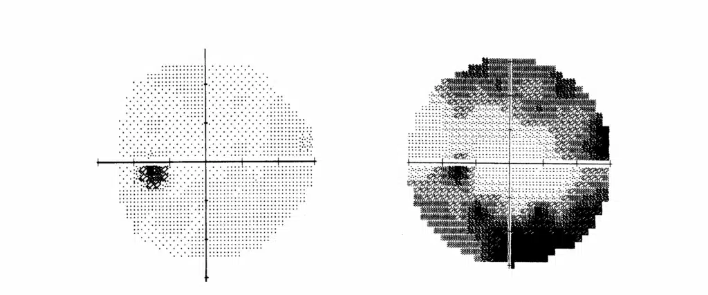
Visual Field Test Results
Results usually include maps, numbers, and reliability markers. The doctor compares these findings with your symptoms, eye exam, and medical history. A single result may not answer every question, so repeat testing may be needed.
Normal and Abnormal Results
Normal results generally show no meaningful missing areas in the tested field. Abnormal results may show reduced sensitivity, missing points, or patterns linked to specific conditions. The location and shape of the defect matter because different diseases affect vision in different ways.
What Makes a Result Reliable?
A reliable result depends on steady focus, clear instructions, and consistent responses. The report may include markers such as fixation losses, false positives, and false negatives. If these markers are high, your doctor may repeat the exam before using the result for diagnosis or treatment planning.
Can You Test Visual Fields at Home?
Home checks can help you notice changes, but they cannot replace formal testing. Tools such as an Amsler grid may help monitor the central area of vision in some patients. They do not measure the full field with the same accuracy as office-based testing.
Limits of Home Vision Checks
Home checks cannot measure light sensitivity, reliability, or subtle field loss as effectively as automated testing. They also cannot show whether the cause is the eye, eyelid, optic nerve, or brain. If you notice a change, a medical exam is the safer next step.
Questions to Ask Your Doctor
- What did my test show?
- Was the result reliable, or should the test be repeated?
- Does the pattern suggest eyelid obstruction, glaucoma, optic nerve disease, or another concern?
- How do these results compare with my symptoms, eye exam, eyelid position, and medical history?
- What should I do next based on the test results?
If you have concerns about changes in your field of vision, drooping eyelids, or abnormal test results, a medical evaluation can help identify the cause and determine the next steps. Dr. Joel Kopelman and the team at Kopelman Aesthetic Surgery can provide a detailed assessment based on your symptoms, eye exam findings, and visual field testing results.